
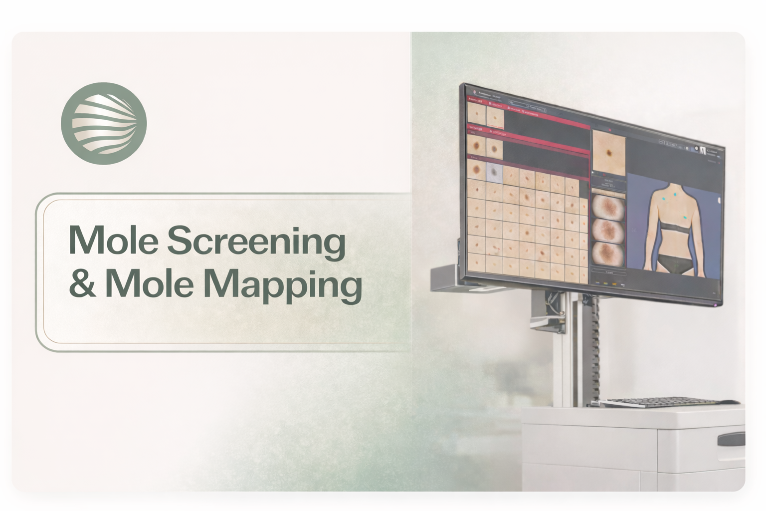
At IA Dermatology, we provide advanced mole screening and full-body mole mapping using state-of-the-art mole scanner AI and digital imaging technology to monitor your skin with clinical precision.
Our consultant dermatologists perform comprehensive skin examinations to identify moles, track changes over time, and provide a vital melanoma check before signs become clinically obvious. This proactive, preventative approach offers patients clarity, reassurance, and long-term skin health monitoring.
This proactive, preventative approach offers patients clarity, reassurance, and long-term skin health monitoring.
Why is a Professional Skin Map Necessary?
Mapping moles allows us to digitally record and compare your skin’s evolution over time. This process helps distinguish harmless spots from pigmented lesions that require further investigation. This is particularly valuable for patients wondering what moles should I get checked, especially those with:
- A high count (over 50 moles).
- A personal or family history of skin cancer.
- Fair skin, red hair, or a history of severe sunburns.
- Atypical or irregular-looking moles.
Every assessment is performed by a senior consultant dermatologist, ensuring clinical judgment guides every decision. If a lesion is found to be suspicious, it can often be addressed through minor dermatologic surgery on the same day.
Who Benefits Most from Mole Screening?
| Lower-Risk Patients | Higher-Risk Patients (Benefit Most) |
| Few moles (under 50). | More than 50 moles on the body. |
| Moles are round, even in colour, and stable. | Irregular or atypical (unusual-looking) moles. |
| No history of skin pigmentation disorders. | Previous abnormal mole or melanoma in the past. |
| Can monitor skin themselves with ease. | Family history of melanoma (parent, sibling, child). |
| Not taking medicines that affect the immune system. | No major sunburn history. |
| Can monitor their skin themselves with ease. | Weakened immune system (e.g., organ transplant patients, long-term immunosuppressive drugs). |
| May choose screening for reassurance. | Fair skin, red hair, freckles, blue/ green eyes with fair skin, history of severe sunburns, especially as a child. |
FotoFinder ATBM: Advanced Mole Scanner AI
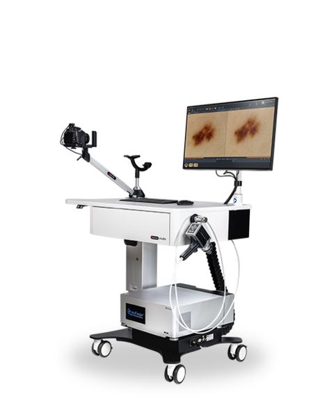
Our clinic utilizes the FotoFinder ATBM (Automated Total Body Mapping) system, the gold standard in melanoma testing technology. Unlike standard photography, this molemap system combines high-resolution imaging with artificial intelligence.
The mole scanner AI automatically locates every mole, allowing for an objective comparison during follow-up visits. This removes the “guesswork” of human memory, making it the most reliable method for checking moles for skin cancer. By detecting the smallest changes early, we can identify cancerous moles in their most treatable stages, often avoiding unnecessary skin treatments or surgeries.
Expert Treatment of Moles
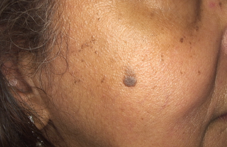
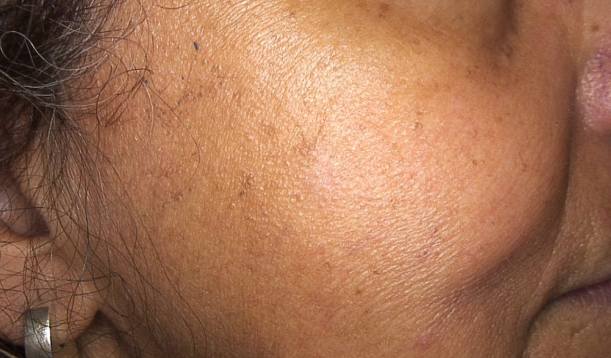
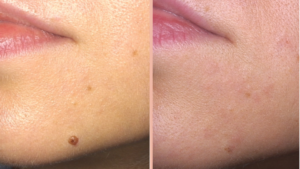
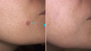
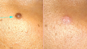
Moles will be surgically removed if they have any suspicious features. This usually involves a simple excision with a very narrow margin (about 1 mm) of normal skin around the mole, designed to minimise scarring. The removed mole is then sent to a specialist laboratory, where it is carefully processed and examined by an experienced pathologist who specialises in skin cancer diagnosis. This ensures the highest quality and accuracy of analysis.
If a mole is confirmed as benign (harmless), no further surgical treatment is required. However, moles may also be removed for cosmetic or practical reasons; for example, if they are catching on clothing, causing discomfort, or if you simply dislike their appearance. In these cases, different techniques may be used, such as flattening a raised mole or reshaping and shrinking a mole to improve cosmetic results. These options would be discussed with you in detail before treatment.
Some mole removal techniques may require a stitch, while others can be performed without stitches. Healing time varies depending on the site: on the face, most mole removals heal within 5–7 days, while on the body, healing typically takes 10–14 days.
Mole Screening & Results
Mole screening is a careful check of your skin, often using a dermatoscope (a special magnifying light) to look closely at moles and other pigmented spots. The aim is to detect skin cancers such as melanoma early, when treatment is most effective.
Benefits of mole screening
✅ Tailored prevention: Provides advice about sun protection and your personal level of risk.
✅ Early detection: Identifies skin cancers at an early stage, often before they become dangerous.
✅ Peace of mind: Reassures you if your moles are harmless.
✅ Accurate monitoring: Photos and digital dermoscopy can track changes over time.
✅ Expert advice: Helps you understand which of your moles are normal and what to watch for in the future.


Spot Changes Early, Stay Confident
Advanced Mole Screening with FotoFinder AI in London
Early detection can save lives. Our painless, AI-powered mole mapping helps identify risks before they become problems.
“Doctor Ali was extremely helpful, understanding and informative and provided excellent care as well as assistance with my future care. It is one of the best doctor-patient experiences I’ve ever had“
✅ Verified Patient Review from Doctify
Visited for Mole Removal
What to Expect During a Mole Screening Consultation
1. Medical History
Your consultation begins with a detailed discussion of your clinical background. Understanding your risk profile is the first step in checking moles for skin cancer. We will discuss:
- Sun Exposure & UV History: Your lifetime history of sunburns and travel to high-altitude or sunny locations.
- Current Medications: Some treatments, such as oral contraceptives for acne or HRT treatments, can influence skin sensitivity or pigmentation.
- Health Background: Conditions like PCOS and hormonal skin or underactive thyroid disease can sometimes impact overall skin health.
- Family History: A history of melanoma or atypical moles in your family significantly influences when to check moles professionally.
2. Comprehensive Skin Examination
You will then have a thorough examination of your skin using a digital dermatoscope.
- This device uses cross-polarised light to provide high-definition analysis of each mole.
- If appropriate, a full-body mole mapping will be undertaken or recommended. This records detailed images of your skin so that moles can be compared over time.
3. Next Steps if a Mole Appears Suspicious
If our melanoma test identifies an unusual lesion, we offer two main clinical paths:
- Monitoring and Review
- If changes are uncertain, the mole will be carefully documented and reviewed again after 4–6 weeks.
- If there are no significant changes, the mole map can continue to be monitored.
- If there are further changes, the mole will be recommended for removal.
- Surgical Excision
- If a mole looks clearly suspicious, removal will be advised.
- This is done under local anaesthetic ( numbing solution injected into the skin with a very fine needle). The surgical procedure usually takes 30–40 minutes.
- A small injection numbs the skin, the mole is removed with a narrow 1 mm margin, and stitches are used to close the area.
- Stitches are removed after 5–7 days on the face or 10–14 days on the body.
- The mole is sent to a specialist laboratory where a pathologist with expertise in skin cancer examines it.
4. Follow-Up and Results
- You will return to the clinic to have your stitches removed and to discuss your results.
- For most patients, no further treatment is required once the mole is confirmed to be benign.
- In a small number of cases, if cancer is diagnosed, further treatment or surgery may be required. This will always be explained in detail and managed appropriately.







FAQs
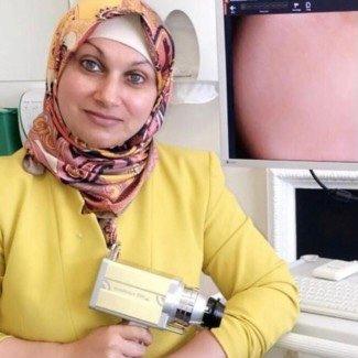
Dr Iaisha Ali, MB ChB MRCP MSc(Oxon)
A London-based dermatologist, specialises in acne, hair loss, hormonal skin disorders, and skin cancer treatment.
Dr Ali has been practising dermatology since 2002, with over 25 years of medical experience delivering consultant-led dermatological care. She previously served as Consultant Dermatologist and Clinical Head of Dermatology at Imperial College Healthcare NHS Trust, where she led complex dermatology services and specialist referral clinics.
